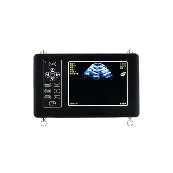
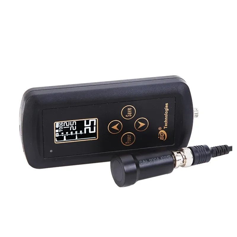
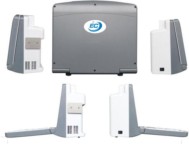
EC-68 Color Doppler Ultrasound Scanner
Product Description
EC68 Full Digital Color Doppler ultrasound Diagnostic System adopts the core technology of
United States. It is a kind of ultrasound imaging system based on PC and ultrasound front-end
combined with kinds of advanced image processing technology at home and abroad. EC68 Full Digital Color Doppler ultrasound Diagnostic System is not only applied to kinds of
general ultrasonic diagnosis as black and white ultrasound system, it is also applied to the
diagnosis with high image quality as CVD, and so on. Functionally, it is equipped with the functions of image scanning, measurement, calculation, display, query, body mark, annotate, printing, medical records storage, editing the inspection
report, system settings, etc. It supports DICOM (Digital Imaging and Communication in
Medicine) protocol, which is a medical imaging standard widely accepted by the world. Data
and information communication can be easily accessed. It also can connect to PACS(Picture
Archiving and Communication System)as a terminal. PACS is a medical picture archiving and
communication systems, which greatly facilitates network management system of the hospital.
It is convenient for remote diagnosis. The machine is powerful, convenient and easy to
operate, and it supports B, B/B, B/4B, B/M, B/PWD, CFM, CDE, B/CFM/D. Meanwhile, the displayer can be moved from up and down, front and back, left and right, that
is really convenient.
Technical Parameters
| No. | Item | Index |
| < 1> | Depth | ≥300mm |
| <2> | Lateral Resolution | ≤ 1mm (Depth≤80mm)≤2mm (80< Depth≤130mm) |
| <3> | Axial Resolution | ≤ 1mm (Depth≤80mm)≤2mm (80< Depth≤130mm) |
| <4> | Blind Area | ≤5 mm |
| <5> | Geometry Position Precision | horizontal≤10%vertical≤10% |
| <6> | Language | English/Chinese |
| <7> | Channels | 32 |
| <8> | Displayer | 12” LCD |
| <9> | External Display | PAL, VGA, |
| <10> | Gray Scale | 256 levels |
| <11> | Voltage | AC220V ± 10% |
| <12> | Operating System | Windows 7 |
| <13> | Scanning Mode | B, B/B, 4B, B/M, M, B+C, B+D, B+C+D, PDI, CF, PW |
| <14> | Probe | Probe sockets: 2Probe frequency: 2 . 0 MHz ~ 1 0 . 0 MHz, 8 -step frequency conversion |
| <15> | Adjustment parameters of color blood flow image | Doppler frequency, sampling frame position and size, baseline, color gain, deflection angle, wall filtering, cumulative times, etc |
| < 16> | Signal processing | With dynamic filtering and quadraturedemodulation With total gain adjustment Gain adjustment: 8-segment TGC The total gain of Type B, Type C and Type D can be adjusted respectively B/ W image gain and color blood flow gain are adjustable respectively |
| < 17> | Doppler | Doppler baseline adjustment level 6Pulse repetition frequency can be adjustedseparately: CFM PWDW ith D linear speed regulation |
| < 18> | Digital beam forming | Continuous dynamic focusing of digital beam forming image Full range dynamic aperture of imageDynamic tracing of the whole image Weighted Sum of Image Whole Process Receiving Delay Support half step scanning and ± 10 ° linear receiving deflection angle Multi beam parallel processing technology |
| < 19> | Basic measurement and calculation function |
Basic measurement in modeB: distance, angle, perimeter and area, volume, stenosis rate, histogram, cross- section |
| Basic measurement of M- mode: heart rate, time, distance and speed | ||
| Doppler measurement: time, heart rate, speed, acceleration | ||
| <20> | Gynecologicalmeasurement andcalculation function | Measurement and calculation of uterus, left ovary, right ovary, left follicle, right follicle, etc |
| <21> | Obstetric measurementand calculation function | G.A, EDD, BPD-FW, FL, AC, HC,CRL, AD, GS, LMP,HL,LV,OFD |
| <22> | Urology measurementand calculation function | Measurement and calculation of left kidney, right kidney, bladder, residual urine volume, prostate, prostate specific antigen predicted value PPSA, prostate specific antigen density PSAD, etc |
| <23> | Product Size | 289×304×222mm |
| <24> | Carton Size | 395×300×410mm |
| <25> | N.W./ G.W. | 6kg/ 7kg |
Standard Configuration
One Host Machine
One Probe Holder
One Convex Array Probe
One Power Adapter
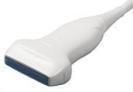
Probe Optional
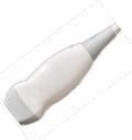
| Probe | C3 - 1/ 60R/3.5MHz Convex Probe | L3 - 1/7.5MHz Liner Probe |
C1 - 6/20R/5.0MHz Micro Convex Probe | EC1 - 1/13R/6.5MHz Transvaginal Probe |
| Picture | 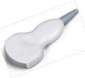 |
 |
 |
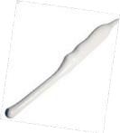 |
| Element s | 80 | 80 | 80 | 80 |
| Scan Width | R60 | L40 | R20 | R13 |
| Frequency | 2 .0/ 3 .0/ 3 .5/4 .0/ 5 .5 MHz | 6 .0/ 6 .5/ 7 .5/ 10/ 12 MHz | 4 .5/ 5 .0/ 5 .5 MHz | 5 . 0/6 .0/6 .5/ 7 .5/ 9 .0 MHz |
| Display Depth | Adjusta ble | Adjusta ble | Adjusta ble | Adjusta ble |
| Scan Depth (mm) | ≧ 160 | ≧50 | ≧80 | ≧40 |
| Resolution Lateral | ≦3 (depth≦80)≦4 (80<depth≦130) | ≦2 (depth≦40 ) | ≦2 (depth≦40 ) | ≦2 (depth≦30 ) |
| Resolution Axial | ≦2 (depth≦80)≦3 (80<depth≦130) | ≦ 1(depth≦4 0 ) | ≦1(depth≦40 ) | ≦1(depth≦40) |
| Blind Area ( mm) | ≦5 | ≦3 | ≦5 | ≦4 |
| Geometric Position (%) Horizontal | ≦15 | ≦ 10 | ≦20 | ≦10 |
| Geometric Position (%) Vertical | ≦10 | ≦5 | ≦10 | ≦5 |